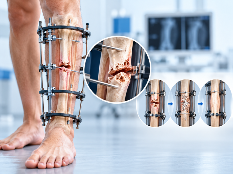
When a fracture is not healing as expected, doctors need to understand exactly what is happening at the break before deciding on another treatment.
Standard X-rays are usually the first imaging test because they can show alignment, fracture lines, callus formation, implants, and changes in healing over time. But some nonunion cases are more complicated. Metal hardware may cover part of the fracture, the break may run at an unusual angle, or it may be difficult to tell how much bone has actually joined.
In these situations, a CT scan can give the orthopedic surgeon a more detailed view before planning nonunion fracture surgery.
Why X-Rays May Not Always Tell the Whole Story
Fracture healing is often monitored using repeated X-rays.
Doctors compare earlier and newer films to look for bridging bone and narrowing of the fracture line. In many cases, this provides enough information to make treatment decisions.
However, an X-ray is a two-dimensional image of a three-dimensional structure.
A plate, rod, or other implant can obscure the fracture. Bone may appear united from one angle but still contain an unhealed area that is difficult to see.
Current reviews of nonunion imaging note that CT is particularly helpful when conventional radiographs are inconclusive or when metallic implants make the fracture site difficult to evaluate.
CT Shows the Fracture From Multiple Angles
A CT scan produces cross-sectional images of the bone.
These images can be reconstructed in different planes, allowing the surgeon to examine the fracture site from angles that are not available on ordinary X-rays.
This can help answer practical surgical questions:
How much of the fracture has actually bridged?
Is there still a clear gap?
Are the bone ends making good contact?
Is part of the fracture healed while another part remains open?
A recent review of long-bone nonunion notes that CT can provide more precise assessment of cortical continuity and bridging callus, particularly in complex fractures.
Understanding the Bone Gap Matters
Some nonunion fractures have very little separation between the bone ends.
Others may have a significant defect because of severe trauma, infection, previous surgery, or loss of damaged bone.
The size and shape of that gap can influence the operation.
A smaller nonunion may mainly need better stability or biological stimulation. A larger defect may require bone grafting or more complex reconstruction.
For patients researching these treatment options, Height Increase Info describes CT scanning as part of comprehensive nonunion assessment alongside X-rays, clinical examination, blood investigations, and other tests when needed.
CT Can Help Identify Alignment Problems
Nonunion is not always just a failure of bone healing.
The fracture may also have healed partly in a poor position or developed rotational or angular deformity.
This can affect walking, joint loading, limb alignment, or the ability to place new fixation correctly.
CT can be especially useful for assessing rotational deformity when the fracture site is difficult to visualize on standard films.
If revision surgery is planned, understanding the three-dimensional alignment beforehand can help the surgeon decide whether the bone needs to be realigned in addition to being stabilized.
Previous Implants Need to Be Evaluated Too
Many patients with nonunion have already undergone fracture surgery.
They may have a plate, screws, an intramedullary nail, or other fixation hardware in place.
Before revision surgery, the orthopedic team needs to understand how that implant relates to the fracture.
Questions may include:
Is the implant still in the correct position?
Has fixation loosened?
Is there enough bone around the existing hardware?
Will the old implant need to be removed?
Where can new fixation safely be placed?
CT can provide additional detail when hardware or overlapping structures make these questions difficult to answer on standard X-rays. Reviews of nonunion imaging also describe CT as useful for assessing bone defects and implant integrity before revision procedures.
CT Can Help With Bone Graft Planning
Bone grafting is commonly considered when a nonunion needs extra biological support or when there is a defect between the bone ends.
The Height Increase Info treatment page lists autograft and allograft among surgical options for nonunion, together with internal fixation, realignment, and infection management.
CT can help the surgeon understand the size and location of the defect before surgery.
That does not determine the graft choice by itself, but it can contribute to planning how much reconstruction may be required and whether stronger fixation is also needed.
A CT Scan Does Not Replace the Rest of the Evaluation
CT is useful, but it should not be treated as the only test needed for nonunion.
Clinical examination remains important.
Doctors also need to understand pain, instability, previous surgery, wound history, and general health. Blood tests may be needed if infection or metabolic problems are suspected.
Importantly, CT also has limitations. Orthopedic consensus literature notes that CT findings can sometimes be difficult to interpret and should be considered alongside ordinary X-rays and the clinical picture rather than used automatically in every case.
This is why CT is usually most helpful when it answers a specific question that standard imaging has not resolved.
How CT Information Changes the Surgical Plan
Once the surgeon understands the nonunion more clearly, the operation can be planned around the actual problem.
Depending on the findings, treatment might involve:
- replacing or strengthening fixation,
- correcting alignment,
- removing unhealthy bone,
- filling a bone defect with graft,
- addressing infection,
- or combining several of these approaches.
The goal is not simply to make the X-ray look better.
It is to create a stable, biologically favorable environment where the bone has another opportunity to unite.
Final Thoughts
CT scans can be valuable when planning nonunion fracture surgery in India because they provide a detailed three-dimensional view of a problem that may be difficult to understand on ordinary X-rays alone.
They can help show persistent fracture gaps, bridging bone, rotational deformity, bone loss, implant position, and other details that may influence revision surgery.
But CT is only one part of the assessment.
The best treatment plan combines imaging with clinical examination, medical history, infection screening, previous surgical information, and an understanding of why the fracture failed to heal in the first place.
In complex nonunion cases, better imaging does not replace surgical judgment.
It gives the surgeon better information on which to base it.